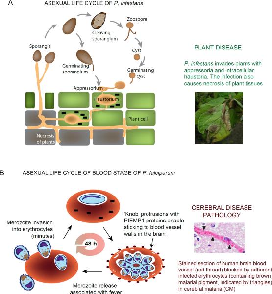
Figure 2. Life cycles and pathologies of Phytophthora and Plasmodium.
(a) P. infestans is able to initiate an infection with both sporangia and zoospores. The pathogen forms an appressorium and penetrates plant leaf cells. Subsequently, a haustorium is developed within host cytoplasm for nutrient acquisition and host immunity modulation. Effectors (black bricks) are trafficked into host cells from the haustoria. Later during infection, P. infestans causes necrosis in plant tissue and sporulates to form new sporangia. The photo of infected potato leaves with sporulating P. infestans is kindly provided by Klaas Bouwmeester and Francine Govers. (b) Asexual, P. falciparum blood stage infection begins when the extracellular merozoite invades the erythrocyte, a process which although complex, is completed in minutes. Effectors (black bricks) are exported across the vacuolar membrane that surrounds the intracellular parasite. Subsequent parasite development, maturation and emergence of merozoites takes ~48 h. In the second half of the asexual cycle, exported parasite effectors assemble in electron dense `knobs' that display the cell surface parasite protein PfEMP1. PfEMP1s are endothelial adhesins that enable infected erythrocytes to bind to the walls of blood vessels. In the brain this can lead to inflammation as well as occlusion of blood vessels (as seen in the adjacent tissue section) and the fatal, neurological pathology of cerebral malaria. The photo is kindly provided by Dan Millner.

