Figure 5.
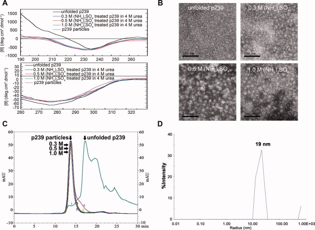
Structural analysis of the (NH4)2SO4-treated p239 in urea. The far-UV and near-UV CD spectra (A) of the p239 particles (purple line), the unfolded p239 (black line), the 0.3M (blue line), 0.5M (red line), and 1.0M (NH4)2SO4-treated p239 in urea (green line) were compared. The EM observation of the unfolded p239, the 0.3M, 0.5M, and 1.0M (NH4)2SO4-treated p239 in urea were studied as labeled (B). The gel filtration profiles of p239 (C) following different treatments indicated the particle formation: unfolded p239 (1, olive green line, equilibration buffer: 10 mM PB7.5 + 4M urea), 0.3M (NH4)2SO4-treated p239 in urea [2, light-green line, equilibration buffer: 10 mM PB7.5 + 4M urea + 0.3M (NH4)2SO4], 0.5M (NH4)2SO4-treated p239 in urea [3, brown line, equilibration buffer: 10 mM PB7.5 + 4M urea + 0.5M (NH4)2SO4], 0.5M (NH4)2SO4-treated p239 in urea [4, magenta line, equilibration buffer: 10 mM PB7.5 + 4M urea + 1.0M (NH4)2SO4], and p239 particles in PBS (5, blue line, equilibration buffer: PBS). mAU, milliabsorbance units. DLS also demonstrated that the 0.3M (NH4)2SO4-treated p239 in urea formed particles with a radius of approximately 19 nm (D). [Color figure can be viewed in the online issue, which is available at wileyonlinelibrary.com.]

