Fig. 7.
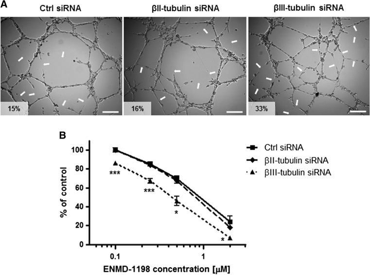
Impact of βII- and βIII-tubulin knockdown on the sensitivity of endothelial cells to vascular-disrupting agent ENMD-1198. a Representative photographs of siRNA-transfected HMEC-1 cells in vascular-disruption assay. Cells were first allowed to form vascular structures on Matrigel for 6 h before drug treatment was initiated. Cells were then incubated for 2 h in presence of ENMD-1198 at 0.25 μM and vascular structures were imaged on a Zeiss Axiovert 200 M using a 5X objective. Arrows point to collapsing and regressing vascular structures. Percentage of vascular disruption is indicated; Scale bar 250 μm. b Dose-dependent effect of ENMD-1198 on the disruption of capillary-like structures formed by siRNA-transfected HMEC-1 cells, after 2 h drug incubation. Points % of intact vascular structures as compared to untreated control cells, means of at least three individual experiments, bars SE; log scale for x axis. Statistics were calculated by comparing the mean surface occupied by closed vascular structures per view field (at least 10 view fields per condition); *p < 0.05; ***p < 0.001

