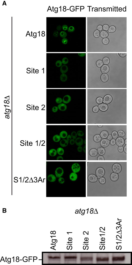
Figure 6. Both phosphoinositide binding sites are required for the proper localization of Atg18 in vivo.
- atg18Δ S. cerevisiae cells transformed with ATG18-GFP, ATG18Site1-GFP, ATG18Site2-GFP, ATG18Site1/Site2-GFP and ATG18Site1/Site2/Δ3Ar-GFP were grown to mid log phase and visualized by confocal microscopy. Representative cells are shown with Atg18-GFP fluorescence images on the left and transmitted light images on the right.
- Western blot analysis was performed using anti-GFP antibodies to determine the overall expression of each Atg18-GFP variant.

