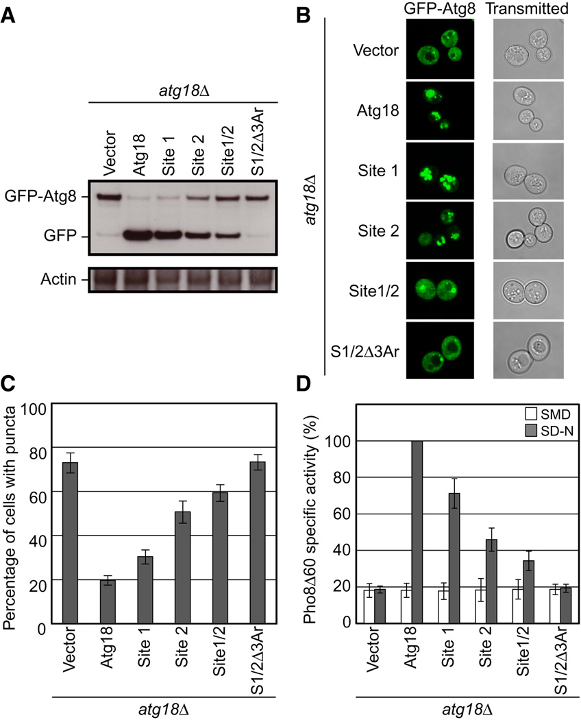
Figure 7. Sites 1 and 2 and the blade 6 loop aromatic residues are important for autophagy.
- GFP-Atg8 processing monitored by western blot against GFP. The full length GFP-Atg8 and GFP bands are labeled. The actin loading control is shown below.
- Representative microscopy images for GFP-Atg8 shown on the left and transmitted light images shown on the right.
- The percentage of cells displaying GFP-Atg8 puncta with a total of 100 cells counted.
- Pho8Δ60 alkaline phosphatase specific activity normalized to starved cells expressing Atg18-myc. Data for non-starved cells are shown in white and starved cells are shown in gray. Error bars in (C) and (D) represent the standard deviation of triplicate experiments.

