
Figure 6.
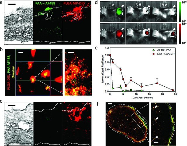
Composite DiD-loaded PLGA microparticle/AF488-loaded PAA microneedles were applied to the flank or dorsal ear skin of C57Bl/6 mice for 5 min, followed by histological, confocal microscope, and whole-animal fluorescence analysis at 1–18 days post treatment. a) Histological section of treated skin 24 h following microneedle array application. Microparticles are shown implanted together with AF488-loaded PAA at a single microneedle penetration site (scale bar 200 μm). b) Parallel x-y/x-z/y-z confocal reconstruction of the treatment site at 24 h post application shows DiD-loaded microparticle depots persisting at the penetration sites (left, scale bar 100 μm). High magnification imaging at a single penetration site shows microparticle dispersion within the cutaneous tissue (right, scale bar 20 μm). c) Histological section of skin 10 days following microneedle application (scale bar 200 μm). d) Whole animal fluorescence imaging of mice 1, 5, and 18 days after microneedle array application. Fluorescence signal from released AF488 and DiD-PLGA microparticles is shown. e) Quantification of relative AF488 and DiD whole-animal fluorescence signal detected at microneedle application sites. f) Histological section of the draining inguinal lymph node 10 days after microneedle application showing persistence of DiD PLGA-loaded cargos in the subcapsular sinus (left, scale bar 100 μm), as well as cell-trafficked PLGA particles in the cortical regions (arrows in inset at right, scale bar 10 μm).