
Fig. 7.
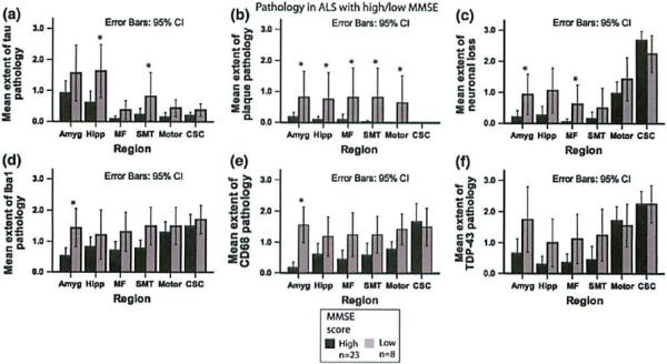
Pathology in ALS related to MMSB. Bar plots Shaw extern of tau (a) and Aβ putholugy (b), as well as ncuronal loss (c), microglial activation (d, e) and TDP-43 patholopy (f) in ALS patients with MMSE above or equal to the median (MMSE high) and MMSE below the median (MMSE low). Whiskers in bar plot indicate 95% confidence Interval of the mean