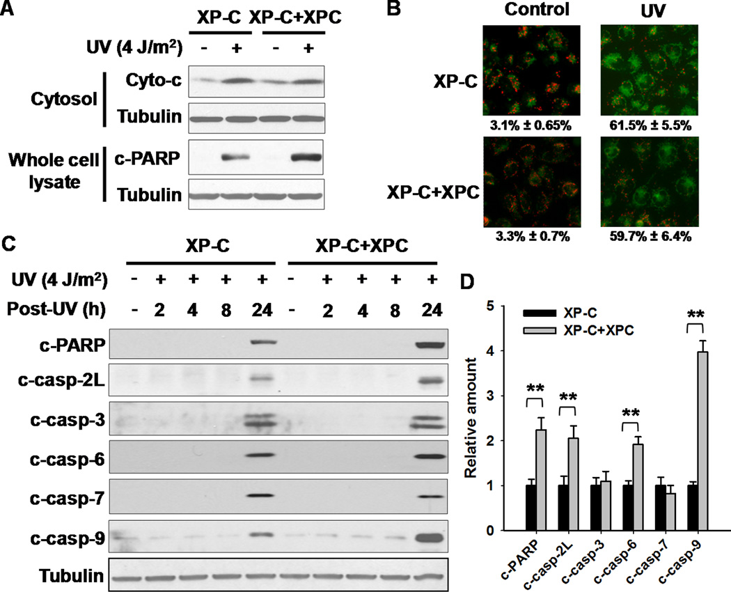
Figure 3.
XPC Functions Downstream of Mitochondrial Events in Facilitating Apoptosis. A, Cytosol was isolated from both XP-C and XP-C+XPC cells at 24 h after UV irradiation. The presence of cyto c in cytosol and the cleaved PARP in whole cell lysates were detected using Western blotting. B, XP-C and XP-C+XPC cells growing on the coverslips were UV irradiated at 4 J/m2, further cultured for 24 h. Mitochondrial trans-membrane potential was detected using MitoCapture probe under fluorescence microscope. The percentages of cells with diffused green fluorescence (apoptotic cells) were calculated. C, XP-C and XP-C+XPC cells were UV irradiated at 4 J/m2, and further cultured for 24 h. Whole cell lysates were prepared and subjected to Western blotting for the detection of cleaved PARP, and various caspases cleavage. D, Quantification of cleaved PARP and various caspases at 24 h after UV irradiation. N = 3, bars: SD, **: p< 0.01, compared with XP-C cells.

