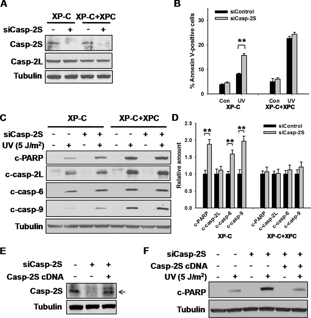
Figure 5.
Casp-2S is Responsible for the Resistance to Apoptosis in XP-C cells. A, XP-C and XP-C+XPC cells were transfected with either control or casp-2S siRNA for 48 h, the expression of casp-2S and casp-2L were detected using Western blotting. B, XP-C and XP-C+XPC cells were transfected with either control or casp-2S siRNA for 48 h, UV irradiated at 5 J/m2 and further cultured for 24 h. Apoptotic cells were detected with Annexin V staining by using Flow cytometry. N = 3, bars: SD, **: p< 0.01, compared with siControl transfected cells. C, Whole cell lysates from cells in (B) were prepared and the cleavages of PARP and various caspases were detected using Western blotting. D, Quantification of cleaved PARP and cleaved caspases after UV irradiation in (C). N = 3, bars: SD, **: p< 0.01, compared with siControl transfected cells. E, XP-C cells were transfected with either casp-2S siRNA, or casp-2S siRNA together with casp-2S cDNA for 48 h, the expression of casp-2S was detected using Western blotting with anti-casp-2S antibody. The Arrow indicates the casp-2S band, the upper band in the third lane is a variant of casp-2S(21). F, Cells in (E) were UV irradiated and further cultured for 24 h. The amount of cleaved PARP and Tubulin was detected using Western blotting.

