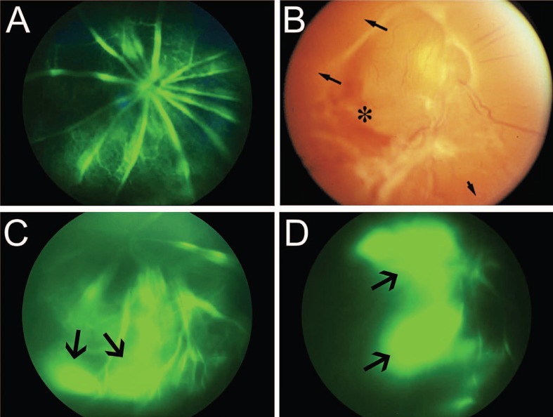
Figure 3.
Defective vascular integrity in laminin null retinas and advanced stage 4 retinopathy of prematurity (ROP). *(A) Fluorescein angiography in wild type P15 mouse retina shows no signs of vascular leakage approximately 120 sec after intra-peritoneal injection of sodium fluorescein. **(B) Advanced stage 4 ROP reveals hemorrhage (asterisk) and avascular peripheral retina (black arrows). *(C) Fluorescein angiography in Lamb2 null retina shows vascular leakage approximately 120 sec after intra-peritoneal injection of sodium fluorescein (black arrow). *(D) Fluorescein angiography in Lamb2:c3 null retina shows vascular leakage approximately 120 sec after intra-peritoneal injection of sodium fluorescein (black arrows). *[Data in A, C and D are from the author’s laboratory]. **[Reprinted with permission from the International Journal of Developmental Biology. Saint-Geniez and D‘Amore, 2004. Originally published in Int J Dev Biol; 48:1045-1058]61

