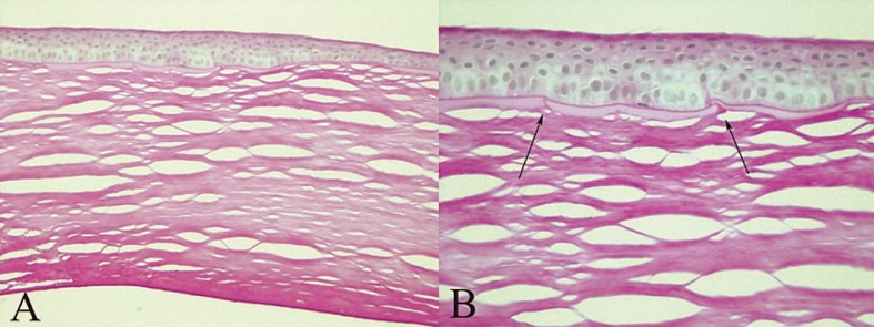
Figure 3.
Histopathology of the graft at the site of ectasia. A, Note focal thickening and irregularity of the epithelium overlying breaks in Bowman’s layer and stromal thinning; the residual recipient stroma, Descemet’s membrane and endothelium are unremarkable (hematoxylin and eosin, ×200); B, Higher magnification of breaks (arrows) in Bowman’s layer (periodic acid-Schiff, ×400).

