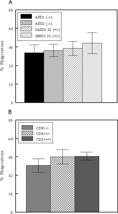
Figure 4.
Phagocytosis by macrophage cell lines. A) ASD1, ASD2, 2ASD1.10, and 2BSD1.10 macrophage cell lines were assessed for phagocytosis of fluorescent beads using flow cytometry. B) Thioglycollate-elicited peritoneal macrophages from CD81−/−, CD81+/− and CD81+/+ mice were assessed for phagocytosis as above. Numbers represent mean ± SEM of four independent experiments.

