
Fig. 7.
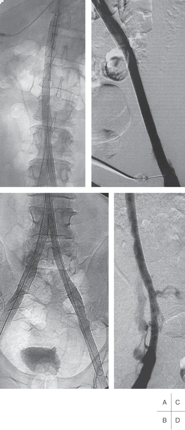
Venograms of the same patient as Fig. 6.
A and B: The chronic occlusion of the IVC has been recanalized and stented from above the renal veins to the right common iliac vein and on the left side to the common femoral vein.
C and D: The stented vein segments of the IVC, and the left and the right iliac veins, respectively, are patent at 1-year follow-up.