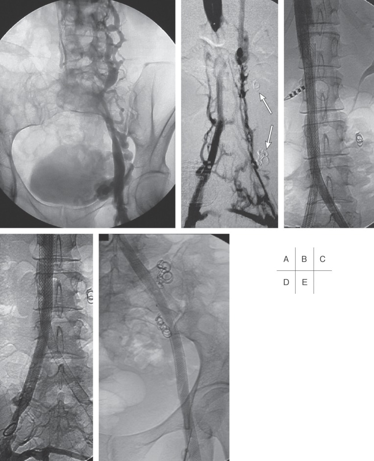
Fig. 9.
Venograms of a 35-year old woman with a chronic occlusion of the IVC following resection of a Wilms tumor of the right kidney in early childhood.
A: Collateralisation via lumbar and ovarian veins.
B: Patent IVC at the level of the left renal vein. Note: coils previously placed in the insufficient left ovarian vein (arrows).
C–E: Unimpeded flow 1 year after recanalization with stent placement from the IVC to the right common iliac vein and to the left common femoral vein.

