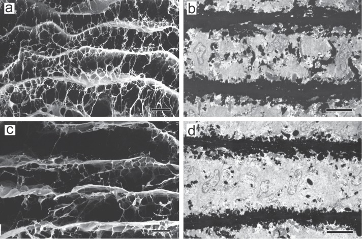
Fig. 5.
The architecture of elastic fibers in experimental aortic dissection. a & b: Normal rat. c & d: β-aminopropionitrile-treated rat. a & c, The aorta was treated with formic acid to remove the components other than the elastic fibers, and then examined by scanning electron microscopy. Scale bar = 20 µm. b & d, The aorta was examined by transmission electron microscopy after tannic acid staining. Scale bar = 5 µm. Upper, intimal side. Lower, adventitial side (Reproduced from Ref. 20).

