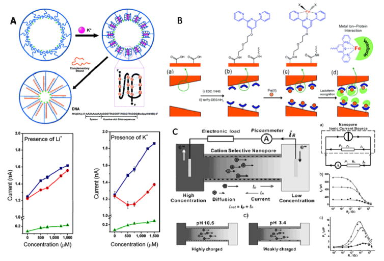
Figure 10. Examples of biomimetic application of solid-state nanopores.
(A) Top: Schematic representation of G4 DNA undergoing conformational change in the presence of K+, and after adding the complementary DNA, a closely packed double-strand DNA was formed, all processes are shown within the nanopore; Bottom: Current-concentration (I-C) characters of the single-conical PET nanopore before and after the G4 DNA molecule modified onto the nanopore inner space. (B) Schematic representation of nanopore-based lactoferrin sensor via the biorecognition of metal-chelating ligand. (C) Left: concentration gradient caused ion diffusion across the cation-selective nanopore; Right: supplying the electrical power produced by the nanopore system to an electric resistance RL, three lines represent different concentration gradient respectively. Figures reproduced with permissions from: (A) Ref. [148], © American Chemical Society; (B) Ref. [149], © American Chemical Society; (C) Ref. [150] © WILEY-VCH Verlag GmbH & Co.KGaA.

