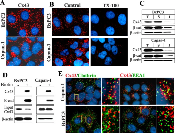
FIGURE 1:
Cx43 fails to assemble into gap junctions in BxPC3 and Capan-1 cells. (A) Cells were immunostained for Cx43. Note that in both BxPC3 and Capan-1 cells, Cx43 (red) is seen as discrete intracellular puncta dispersed throughout the cytoplasm. (B) Loss of Cx43 intracellular puncta upon in situ extraction with 1% TX100 in BxPC3 and Capan-1 cells. Note loss of Cx43 intracellular puncta upon extraction with TX100. (C) Western blot analysis of Cx43 in total (T), TX100-soluble (S), and TX100-insoluble (I) fractions of cell lysates in BxPC3 and Capan-1 cells. Note that the majority of Cx43 is soluble in TX100 in both cell types. (D) Cx43 traffics to the cell surface in BxPC3 and Capan-1 cells. The cell surface proteins of BxPC3 and Capan-1 cells were biotinylated. Biotinylated proteins were pulled down by immobilized streptavidin and immunoblotted for Cx43. Biotinylation of E-cadherin (E-cad) was used as a positive control. For input, 10 μg total cell lysate was used. Note that Cx43 and E-cad were efficiently biotinylated in both cell lines. (E) Cells were immunostained for Cx43 (red), clathrin, and EEA1. Enlarged images of the boxed regions are shown on the right. Note that Cx43 does not colocalize discernibly with either marker.

