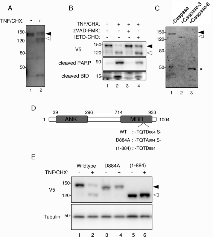
FIGURE 1:
Caspase-3–mediated cleavage of MYPT1 during apoptosis. (A) Cleavage of endogenous MYPT1 in apoptotic cells. HeLa cells were treated with dimethyl sulfoxide (DMSO) or TNF/CHX for 3 h. Total cell lysates were immunoblotted with anti-MYPT1 antibody. (B) Inhibition of MYPT1 cleavage. HeLa cells were transiently transfected with V5-MYPT1-WT. At 24 h after transfection, the cells were treated with DMSO, TNF/CHX, TNF/CHX zVAD-FMK, or TNF/CHX/IETD-CHO for 3 h. Total cell lysates were immunoblotted using anti-V5 antibody (top panel), cleaved PARP antibody (middle panel), or BID antibody (bottom panel). (C) Identification of MYPT1-cleaving caspase. FLAG-MYPT1 was treated with caspase-3 or caspase-8 at 30˚C for 1 h in vitro. Cleavage of MYPT1 was analyzed by immunoblot assay using the anti-FLAG antibody. Asterisk indicates the caspase-8–cleaved form of MYPT1. (D) Schematic representation of MYPT1. ANK, ankyrin repeat; MBD, myosin-binding domain. The caspase-3 cleavage site TQTD884 was identified by amino acid sequencing. D884A and (1-884) indicate the caspase-3 cleavage site mutant and the caspase-3–cleaved form of MYPT1 (lacking the C-terminal end), respectively. (E) In vivo cleavage of MYPT1 at Asp-884. HeLa cells transfected with V5-MYPT1-WT, V5-MYPT1-D884A, or V5-MYPT1-(1-884) were treated with DMSO or TNF/CHX for 3 h. Total cell lysates were immunoblotted using anti-V5 antibody (top panel) or anti–α-tubulin antibody (bottom panel). Positions of the full-length and caspase-3–cleaved MYPT1 are indicated using filled and open arrowheads, respectively. Positions of molecular weight standards are indicated in kilodaltons on the left.

