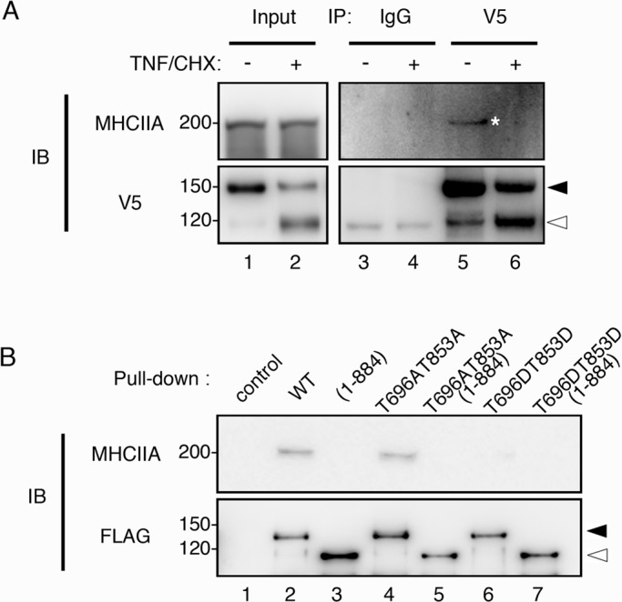
FIGURE 3:

Dissociation of MYPT1 from myosin II during apoptosis. (A) HeLa cells expressing V5-MYPT1-WT were treated with DMSO or TNF/CHX for 3 h. Immunoprecipitates prepared with anti-mouse immunoglobulin G (IgG, lanes 3 and 4) and anti-V5 antibody (V5, lanes 5 and 6), respectively, were immunoblotted with anti–myosin heavy chain IIA antibody (MHCIIA). (B) Pull-down assay from nonapoptotic HeLa cell lysates was performed using FLAG-MYPT1-WT, FLAG-MYPT1-(1-884), FLAG-MYPT1-T696AT853A, FLAG-MYPT1-T696AT853A-(1-884), FLAG-MYPT1-T696DT853D, and FLAG-MYPT1-T696DT853D-(1-884), respectively (lanes 2–7). The precipitates were probed with anti–myosin heavy chain IIA antibody (MHCIIA). Filled and open arrowheads indicate the positions of full-length and caspase-3–cleaved MYPT1, respectively.
