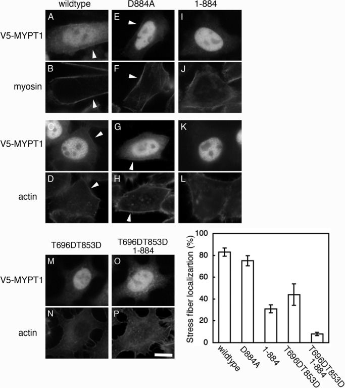
FIGURE 4:

Localization of caspase-3–cleaved MYPT1. HeLa cells transiently transfected with V5-MYPT1-WT (A–D), V5-MYPT1-D884A (E–H), V5-MYPT1-(1-884) (I–L), V5-MYPT1-T696DT853D (M and N), and V5-MYPT1-T696DT853D-(1-884) (O and P) were doubly stained with anti-V5 antibody and either with anti–myosin heavy chain IIA antibody or with phalloidin. Localization of MYPT1 at the cortex region is indicated by white arrowheads. Scale bar: 30 μm. Graph shows percentage of the cells in which V5-MYPT1 is localized at stress fibers. Results shown are mean ± SD of three independent experiments.
