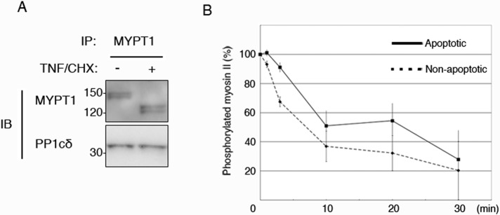
FIGURE 5:

Phosphatase activity of apoptotic MP. MP was immunoprecipitated by anti-MYPT1 antibody from nonapoptotic cells (A, lane 1) and apoptotic cells (A, lane 2). Coimmunoprecipitation of MP component was confirmed by anti-PP1cδ antibody. Myosin II, phosphorylated with ZIPK, was incubated with apoptotic (B, solid line) and nonapoptotic (B, dotted line) MPs for the indicated times. Remaining amount of phosphorylated myosin II was determined using anti–Ser-19 phosphorylated MRLC antibody. Each data point represents mean ± SD of three independent experiments.
