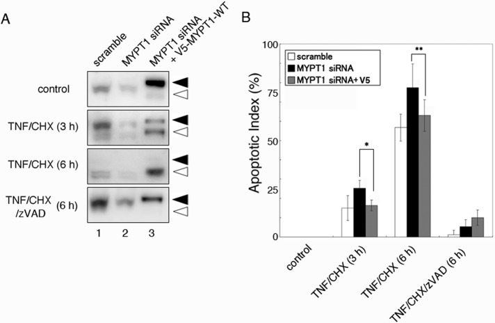
FIGURE 6:
Effect of MYPT1 depletion on apoptotic cells. (A) HeLa cells were transfected with scrambled siRNA (lane 1), human MYPT1 siRNA (lane 2), or human MYPT1 siRNA and V5-MYPT1-WT vector (lane 3). Forty-eight hours after transfection, the cells were treated with DMSO, TNF/CHX, or TNF/CHX/zVAD-FMK for the indicated times. Total cell lysates were immunoblotted with anti-MYPT1 antibody. The full-length and caspase-3–cleaved MYPT1s are indicated using filled and open arrowheads, respectively. (B) The dead cells in the indicated time were counted after staining the cells with FITC-annexin V and PI. The apoptotic index was calculated by determining the percentage of dead cells in the field (n = 100–200). Results shown are mean ± SD of four independent experiments. Statistical significance was determined using Student's t test (*, p < 0.005; **, p < 0.05).

