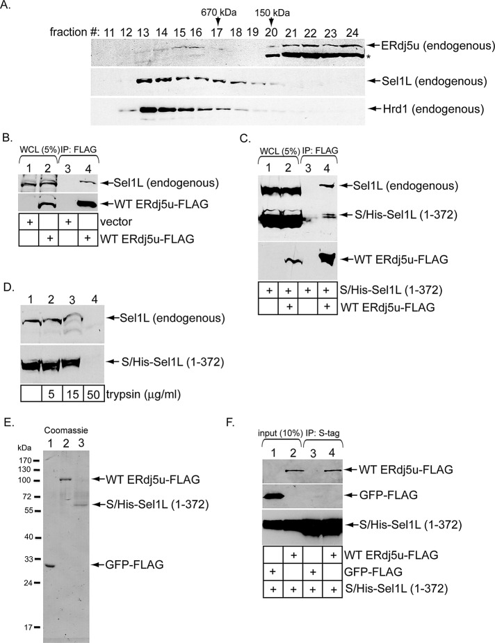
FIGURE 4:
ERdj5u binds to Sel1L. (A) A WCL derived from 293T cells was subjected to gel filtration. Individual fractions were subjected to SDS–PAGE and analyzed by immunoblotting using the indicated antibodies. The asterisk denotes ERp72 that cross-reacts with an ERdj5 antibody (Proteintech Group). (B) Cells were transfected with vector or WT ERdj5u-FLAG and lysed in a buffer containing 1% deoxy Big CHAP. The resulting WCLs were incubated with FLAG antibody–conjugated beads, and the immunoprecipitates were subjected to SDS–PAGE followed by immunoblotting with the indicated antibodies. (C) Cells transfected with S/His-Sel1L (1–372) alone or S/His-Sel1L (1–372) and WT ERdj5u-FLAG were lysed in a buffer containing 1% deoxy Big CHAP. The resulting WCLs were processed as in (B). (D) A WCL expressing S/His-Sel1L (1–372) was incubated with the indicated trypsin concentration, subjected to SDS–PAGE, and immunoblotted using an antibody against Sel1L. (E) GFP-FLAG, WT ERdj5u-FLAG, and S/His-Sel1L (1–372) were expressed in 293T cells, and the purified proteins were subjected to SDS–PAGE followed by Coomassie blue staining. Fraction 27 during purification of S/His-Sel1L (1–372) is shown. (F) S/His-Sel1L (1–372) was incubated with either GFP-FLAG or WT ERdj5u-FLAG, and the samples were precipitated using S-antibody–conjugated beads. The immunoprecipitates were subjected to SDS–PAGE followed by immunoblotting with the indicated antibodies. The input samples were also analyzed by immunoblotting with the appropriate antibodies.

