FIGURE 8:
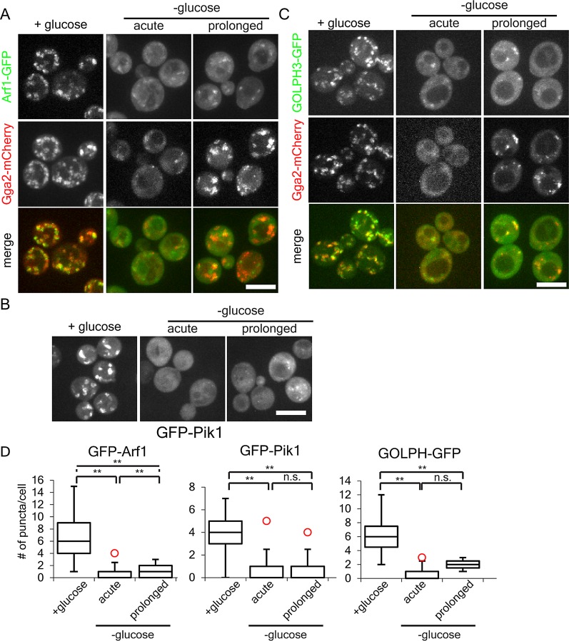
Glucose starvation alters the localization of Arf1, Pik1, and PI4p. (A) Arf relocalizes to dim puncta during acute and prolonged starvation. Diploid cells heterozygous for ARF1-GFP and homozygous for GGA2-mCherry were imaged before, within 15 min, or after 2 h of glucose starvation. (B) Pik1 redistributes to the cytosol upon glucose starvation. Haploid wild-type cells expressing GFP-Pik1 from a plasmid were imaged before, within 15 min, or after 2 h of glucose starvation. (C) GOLPH3 relocalizes to dim puncta during acute and prolonged starvation. Haploid wild-type cells expressing Gga2-mCherry from the endogenous locus and the PI4P probe GOLPH3-GFP from a plasmid were imaged before, within 15 min, or after 2 h of glucose starvation. (D) Quantification of puncta per cell for cells grown in or acutely starved for glucose as described in A–C. **p < 0 .01; n.s., not significant. Charts show box plot of data as described in Figure 1.

