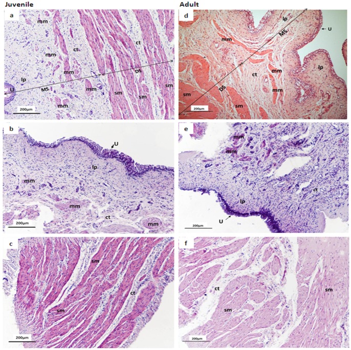
Figure 5. Low-power micrographs showing H&E staining of representative intact, mucosal and denuded-detrusor strips from juvenile and adult pig bladder dome.
Left panel shows representative sections from juvenile bladder intact strip a) and mucosal strip (b) and denuded-detrusor strip (c); right panel shows an intact strip (d), a mucosal strip (e) and a denuded detrusor strip (f) from adult pig bladder. Note that the arrows in sections a) & d) demonstrate the plane of division between the mucosal and denuded-detrusor strips. MS, mucosal strip; DS, denuded-detrusor strip; sm, smooth muscle; ct, connective tissue; lp, lamina propria; U, urothelium; mm, muscularis mucosa.

