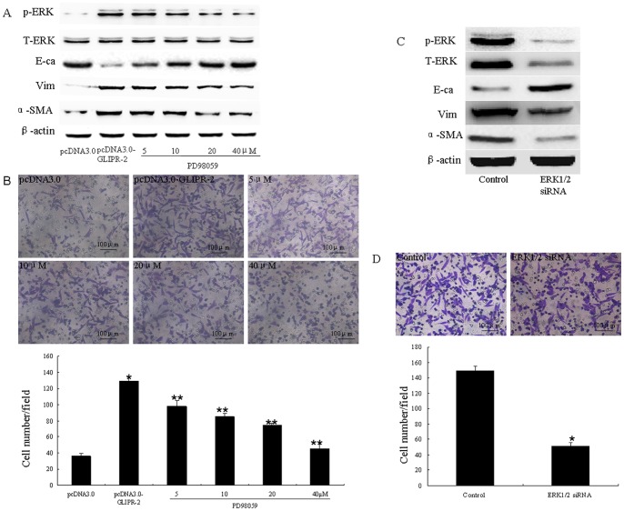
Figure 3. GLIPR-2 overexpression in HK-2 cells promotes an EMT through ERK1/2 activation.
(A)Western blot data of EMT markers and ERK1/2 activation in GLIPR-2-overexpressing HK-2 cells. p-ERK1/2 was elevated in the pCDNA3.0-GLIPR-2-transfected HK-2 cells but decreased gradually in a dose-dependent manner with PD98059 treatment; E-cadherin decreased in the pCDNA3.0-GLIPR-2-transfected HK-2 cells but increased gradually in a dose-dependent manner with PD98059 treatment; vimentin and α-smooth muscle actin increased in the pCDNA3.0- GLIPR-2-transfected HK-2 cells but decreased gradually in a dose-dependent manner with PD98059 treatment.(B) Promotion of cell migration by GLIPR-2 overexpression, and the inhibition of cell migration via the abrogation of ERK1/2 activity with PD98059 in the GLIPR-2-transfected group. Two groups of confluent, growth-arrested, and serum-starved HK-2 cells were seeded onto the upper part of the Millicell (8-µm pore size) with various concentrations of PD98059 in the GLIPR-2-transfected group. After incubating the cells for 8 hrs, the number of migrating cells was counted as described above. The data are presented as the mean ± SD. *P<0.01 compared with the pcDNA3.0 group. **P<0.01 compared with the GLIPR-2-transfected group. One of three individual experiments is shown. (C) Western blot data of EMT markers and ERK1/2 activation in the ERK1/2 siRNA-transfected HK-2 cells that overexpressed GLIPR-2. Total-ERK1/2 and p-ERK1/2 decreased and EMT markers were reversed after treatment with the ERK1/2 siRNA. (D) Inhibition of cell migration by ERK1/2 siRNA transfection of the HK-2 cells which overexpressed GLIPR-2. The data are presented as the mean ± SD. *P<0.01 compared with the control groups.

