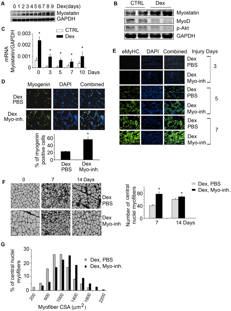
Figure 7. Dex increases myostatin expression and impairs satellite cell activation in vivo.
A. Representative western blots of myostatin in gastrocnemius muscles of mice treated with Dex for different days. B. Representative western blots of indicated proteins from muscles of control or mice treated with Dex for 14 days. C. mRNA expression of myostatin was measured by RT-PCR in muscles of mice treated with or without Dex and injured with CTX (*p<0.05 vs. CTRL; n = 3 mice in each group). D. At 4 days after injury, cross-sections of muscle were immunostained with anti-myogenin (left panel) and the ratio of myogenin positive cells to DAPI expressed as a percentage is shown in right panel (*p<0.05 vs. Dex plus PBS). E. Cross-sections of injured TA muscles from mice injected with Dex plus PBS or Dex plus myostatin inhibitor were immunostained with anti-eMyHC (green). F. Sections in Fig. 7E were immunostained with laminin and DAPI to show the newly formed myofibers (left panel). The average number of central nuclei myofibers was calculated from 10 areas counted (*p<0.05 vs. Dex plus PBS, right panel). G. at 14 days after injury, newly formed myofiber cross-sectional areas were measured and the distribution is shown.

