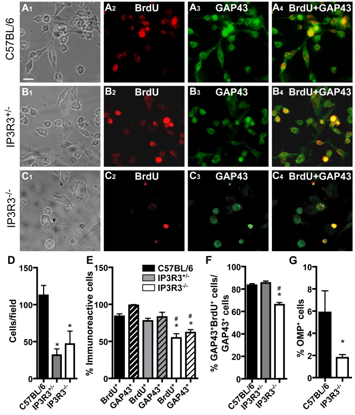
Figure 4. The capacity of basal cells to differentiate is compromised in the OE of IP3R3−/− mice.
(A–C) Representative images of OE explant cultures from neonatal (A) C57BL/6, (B) IP3R3+/− and (C) IP3R3−/− mice exposed to BrdU (50 µg/ml) from day 4–8. Cells surrounding the OE explant immunoreactive to antibodies directed against BrdU (A2–C2), immature neuron marker GAP43 (A3–C3), to both BrdU and GAP43 (A4–C4), or mature neuron marker OMP (data not shown) were quantified. Scale bar = 50 µm. (D) There was a significantly lower number of cells surrounding the explants from IP3R3+/− and IP3R3−/− mice than compared to C57BL/6 mice. *, p<0.05 v. C57BL/6 (One way ANOVA with Newman-Keuls post-hoc test; n = 3, 3 and 5 coverslips, respectively.) Refer to legend in E. (E) BrdU+ and GAP43+ cells in the explant culture from IP3R3−/− mice were significantly lower compared to C57BL/6 and IP3R3+/− mice. *, p<0.01 v. C57BL/6; #, p<0.05 v. IP3R3+/− (Two way ANOVA with Newman-Keuls post-hoc test; n = 3, 4, 3 coverslips, respectively). Legend refers to D, F–G. (F) Co-localization of BrdU+ and GAP43+ cells in the explant culture from IP3R3−/− mice was significantly lower compared to IP3R3+/− and C57BL/6 mice. *, p<0.01 v.C57BL/6, #, p<0.001 v. IP3R3+/− (One way ANOVA with Newman-Keuls post-hoc test; n = 3 coverslips, each.) (G) OMP+ cells in the explant culture from IP3R3−/− mice were significantly lower compared to that of C57BL/6 mice. *, p<0.02 (Student’s t-test; n = 3 and 6 coverslips, respectively).

