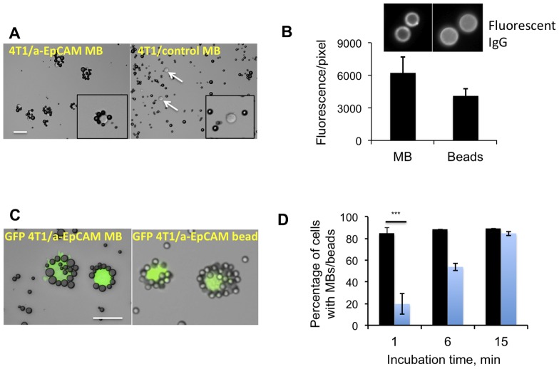
Figure 2. Binding of MBs to cultured tumor cells.
A, Anti-EpCAM MBs and control MBs were added at 100∶1 ratio to a suspension of 100,000 4T1 mouse breast carcinoma cells in 1 ml cell medium and mixed for 15 min. Targeted MBs formed rosettes around the cells, while control MBs did not show any binding. Size bar, 50 µm for both images; B, Magnetic beads (5 µm diameter) were decorated with the anti-EpCAM antibody according to the strategy described in Fig. 1. Anti-EpCAM IgG was detected on MBs and beads using a secondary fluorescent antibody; the coating was comparable for MBs and beads; C, MBs and beads were mixed with GFP-4T1 cells in 1 ml medium (100∶1 ratio). The characteristic rosettes between MBs and beads were observed after 15 min; D, Binding efficiency of MBs and magnetic beads was determined at different time points. Blue bars, percentage of cells coated with beads; black bars, percentage of cells coated with MBs. After 1 min, anti-EpCAM MBs bound to 4T1 cells more efficiently than anti-EpCAM magnetic beads (t-test, P = 0.0001, n = 3).

