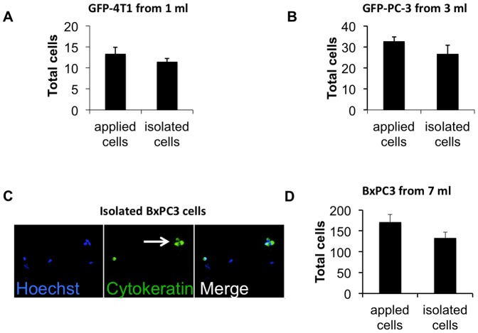
Figure 5. Isolation of rare cells with MBs.
Rare tumor cells were added to plasma-depleted blood, isolated with MBs as described in Fig. 4A and counted on a slide. In order to avoid spiking and counting errors, the same number of tumor cells that was added to blood cells prior to isolation (typically in 5–10 µl volume) was placed on a slide and counted in parallel with the isolated sample; A, Mouse breast cancer GFP-4T1 cells were added to 1 ml blood and isolated with anti-mouse EpCAM MBs (n = 3). B, Prostate cancer GFP-PC-3 cells were added to 3 ml of plasma-depleted blood and isolated with anti-human EpCAM MBs (n = 3); C, Pancreatic cancer BxPC3 cells were added to 7 ml plasma-depleted blood and isolated with anti-human EpCAM MBs. Unlike the experiments with GFP-tagged cells, the isolated cells were stained with pan-cytokeratin antibody. A representative microscopic field (20×objective) shows the MB-isolated BxPC3 cells positive for CK (green). Hoechst-positive, CK-negative cells, which are presumably carryover leukocytes, are also visible in the field. Arrow points to a tumor cell cluster; D, There was a 77% efficiency of isolation of BxPC3 cells from 7 ml (n = 3).

