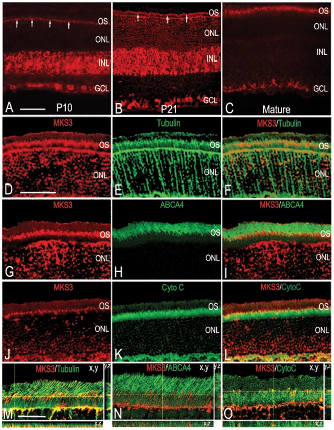
Figure 1. Meckelin 3 in the developing rat retina.
Confocal imaging of wild-type P10 (A), P21 (B), and mature (C) retinal sections immunolabeled with antibody specific for MKS3 showed that MKS3 expression was widespread in the early postnatal retina that becomes restricted in the mature retina. (A) At P10 MKS3 could be detected in the region of the ONL consistent with developing inner and outer segments (arrows), as well as the inner and ganglion cell layers. (B) At P21, MKS3 showed similar localization in comparison to P10, but developing outer segments were much more visible at this stage (arrows). (C) MKS3 label was detected in photoreceptor outer segments and ganglion cells; no label in inner nuclear was detected at this stage. Retinal sections were also double-labeled with MKS3 (D,G, J) and tubulin (E), ABCA4 (H), or cytochrome C (K) to better localize MKS3 protein within the photoreceptor inner and outer segments. An overlay of MKS3 and tubulin (F) highlighted the localization of MKS3 in the photoreceptor axoneme, which spans the inner segment, connective cilium, and the lower portion of the outer segment. Consistent with the localization of MKS3 within the axoneme, there was a partial overlap with cytochrome C (L) within the upper portion of the inner segment. There appears to be little to no overlap with the outer segment marker, ABCA4 (I), indicative of MKS3 localization primarily to the connective cilium and lower portion of the outer segment. Thin plane confocal microscopy on sections labeled for MKS3 and tubulin (M), ABCA4 (N), or Cytochrome C (O) in order to verify patterns of expression. For M, N, and O, the xy, planes are labeled and the yellow lines indicate the x,z and y,z planes depicted in the strips to the right and the bottom. GCL, ganglion cell layer; INL, inner nuclear layer; ONL, outer nuclear layer; OS, outer segment; CytoC, cytochrome C; ABCA4, ATP-binding cassette sub-family A – member 4. Scale bar: (A) Bar = 50 µm for panels A–C, (D) Bar = 50 µm for panels D–L, (M) 100 µm for M–O.

