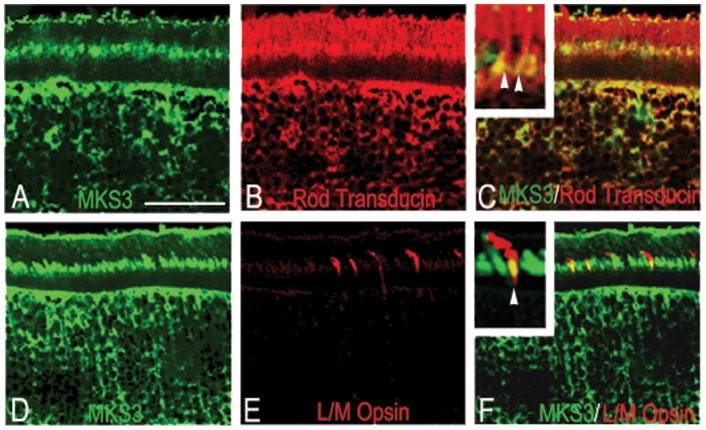
Figure 2. MKS3 was found in rods and cones.
Double-label immunohistochemistry was performed on sections through P21 WT retina using an antibody specific for MKS3 (A, D) and rod transducin (B) or L/M opsin (E). Confocal images were then merged into a single image (C, F) to show the co-expression of MKS3 and photoreceptors in the outer segment. Increased magnification in insets (C, F) indicated there is only partial overlap in localization of both rod transducin and L/M opsin with MKS3. GCL, ganglion cell layer; INL, inner nuclear layer; ONL, outer nuclear layer; OS, outer segment. Scale bar: (A) 50 µm.

