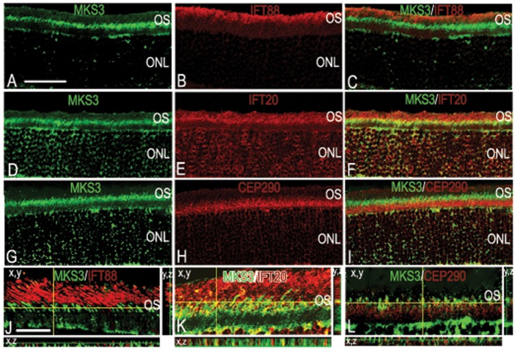
Figure 3. MKS3 showed partial co-localization with IFT20.
To elucidate which IFT proteins might interact with MKS3, we performed double-label immunohistochemistry with MKS3 (A, D, G) and IFT88 (B), IFT20 (E), and Cep290 (H). Overlay of confocal images (C, F, I) showed that MKS3 was partially co-localized with IFT 20 (F). MKS3 was not co-localized with IFT88 (C), or Cep290 (I). Localization data was verified using thin-plane confocal microscopy (J–L). In J–L, the horizontal yellow line in the x,y image indicates the focal plane illustrated in the x,z strip at the bottom of the panel and the vertical yellow line in x,y image indicates the focal plane illustrated in the y,z panel. Scale Bar = (A) 50 µm for panels A-I and (J) 100 µm for panels J–L.

