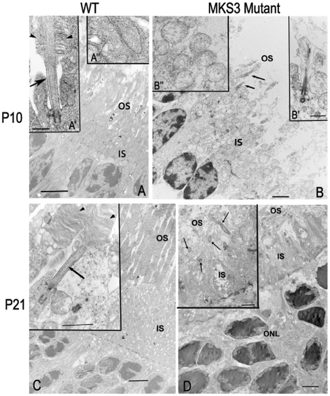
Figure 7. Electron microscopy of photoreceptor outer segments.
Electron microscopic images were compared at P10 and P21 in WT (A, C) and mutant (B, D) retinae. At P10, the nascent outer segment discs can be found in WT (A', arrowhead) while in the mutant (B, arrows) the outer segment was not apparent, but a membranous loop without discs was associated with the cilium. Mitochondria in WT P10 appeared as elongated structures with visible cristae (A’’), while in the mutant retina the mitochondria appeared swollen with no evidence of cristae (B’’). At P21, the WT retina has a well-developed OS with discs (C), while the mutant (D) demonstrates cilia extending into the OS space with some loosely associated membranous material, but no discs were observed (inset, D). (A, C) IS; inner segments, OS; outer segments, WT; wild type. Scale Bar = 4 µm, (B, D) 2 µm; Insets for A = 500 nm, B- D = 1 µm.

