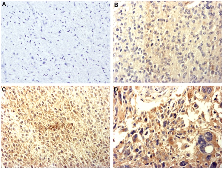
Figure 1. Immunohistochemistry staining pattern of EMMPRIN in glioma (×200). A.
Negative staining (−) of EMMPRIN, figure was taken from grade III glioma; B Weak positive staining (+) of EMMPRIN, figure was taken from grade II glioma; C Moderate positive staining (++) of EMMPRIN, figure was taken from grade II glioma; D Strong positive staining (+++) of EMMPRIN, figure was taken from grade IV glioma.

