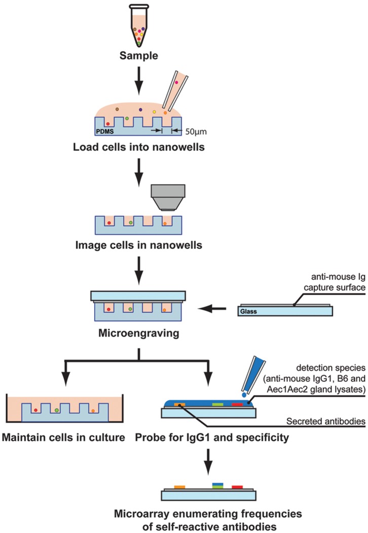
Figure 1. Illustration of microengraving.
Arrays of nanowells with dimensions of 50 µm×50 µm×50 µm were used for microengraving. Spleen or cervical lymph nodes cells were loaded in the nanowells. Cells in the nanowells were imaged using an automated epifluorescence microscope. Micrograving is performed by hybridizing nanowells with capture slides containing anti-mouse Ig for 2 hrs at 37°C with 5% CO2. After incubation, nanowells containing intact live cells and capture slides were separated. A mixture of antibodies containing IgG1-Alexa Fluor 488, B6 SG lysate-Alexa Fluor 594 and Aec1Aec2 SG lysate-Alexa Fluor 555 were added to the capture slides. Micrographs of microarrays were generating by scanning using a Genepix 4200AL microarray scanner.

