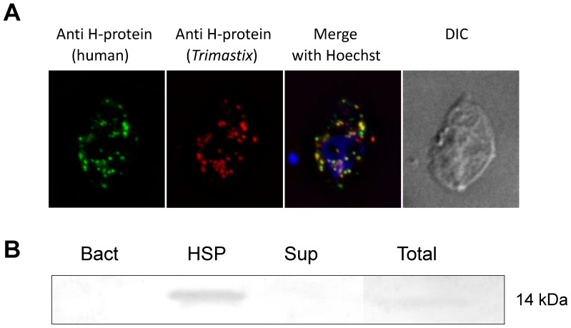
Figure 2. H-protein of GCS localizes into vesicles (putative mitochondrion-like organelles) in Trimastix pyriformis.
A) Immunofluorescence microscopy of the Trimastix pyriformis cell. The green signal from antiH-protein (human) co-localizes with red signal from the antiH-protein (Trimastix). The DNA is stained blue with Hoechst. B) Western blot on the cellular fractions of Trimastix pyriformis. The lines represent pure bacteria Citrobacter sp. from the culture (Bact), high speed pellet of Trimastix (HSP), supernatant of Trimastix (Sup), total lysate of Trimastix (Total).

