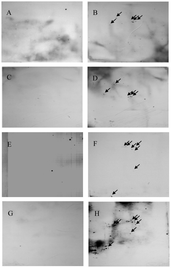
Figure 1. 2DE SDS-PAGE/immunoblot analysis of total cellular proteins of HEK293 cells infected with Ad5 or Ad35.
The total protein of Ad5 (B, D) and Ad35 (F, H) infected and mock infected (A, C, E, G) HEK293 cells were separated by 2DE SDS-PAGE. 2DE SDS-PAGE gels were transferred onto a nitrocellulose membrane. Blots were probed with in-house rhesus monkeys anti-wild Ad5 (A, B) and Ad35 antibodies (E, F) or healthy adult human serum positive for Ad5 (C, D) and Ad35 (G, H), followed by incubation with the corresponding horse radish peroxidase (HRP)-conjugated goat anti monkey or goat anti human IgG secondary antibodies.

