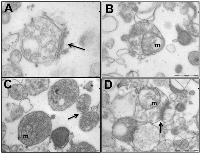
Figure 4. Cerebral Synaptosomal Ultrastructural Abnormalities Were Corrected by Antipurinergic Therapy.
(A) Control (Sal-Sal) synaptosome illustrating normal post-synaptic density (PSD) morphology (arrow), and normal electron lucency of the matrix (92,000× magnification; scale bar = 200 µm). (B) Treated controls (Sal-Sur) with an included mitochondrion (“m”; scale bar = 500 µm). (C) Untreated MIA (PIC-Sal) with an included mitochondrion (“m”) and malformed, hypomorphic PSD (arrow; scale bar = 500 µm). Note the abnormal accumulation of electron-dense matrix material. (D) Treated MIA (PIC-Sur) with restoration of near-normal PSD morphology (arrow), an included mitochondrion (“m”), and reduction in abnormal accumulations of electron-dense matrix material within the synaptosomes (scale bar = 500 µm). Representative fields from n = 3–4 males per group; age = 16 weeks.

