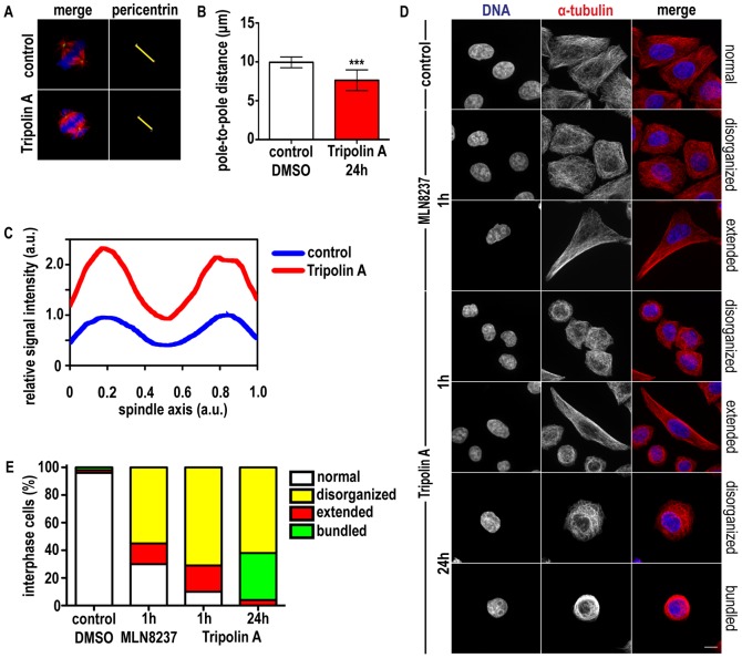
Figure 4. Tripolin A alters pole-to-pole distance and MT stability in mitotic cells and influences interphase MT array.
(A) Maximum projections from z-stacks of a representative control cell and representative cells treated with Tripolin A. In the merged images α-tubulin is pseudocolored red; pericentrin is green, DNA is blue. Yellow arrows indicate interpolar distance. (B) Interpolar distances were measured based on pericentrin staining in HeLa cells (n≥100 cells for each group, from at least three independent experiments). ***: p<0.0001; (Student's t-test, two-tailed). Error bars indicate SD. (C) Longitudinal line scans of tubulin intensity from metaphase spindles of control and Tripolin A treated HeLa cells (n = 5 for each group). Intensities were normalized to the maximum value of the control curve, and spindle size was interpolated. Curves indicate mean values. (D) Representative immunofluorescence images of HeLa cells in interphase treated with DMSO, 100 nM MLN8237 for 1 h or 20 µM Tripolin A for 1 h and 24 h. In the merged images α-tubulin is pseudocolored red, DNA blue. (Scale bar 10 µm). (E) Graph showing the percentages of interphase cells with altered MT array, classified in the indicated arbitrary categories in control cells (DMSO) and cells treated with MLN8237 or Tripolin A (n = 150 cells for each group, from three independent experiments).

