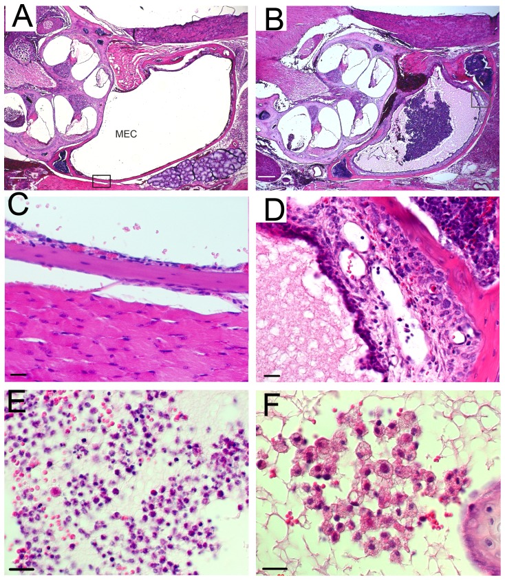
Figure 5. Hematoxylin and eosin staining of the middle ear in adult mice indicated otitis media.
Clear middle ear cavity (MEC) and thin mucoperiosteum in wild type mice (A,C). MECs of Mcph1tm1a /tm1a mice (B,D) were filled with exudate and lined with thickened mucoperiosteum. High magnification for mucoperiosteum of MEC framed in A and B (C,D). Inflammatory cells (E,F) in MECs. Scale bar, 200 µm (A,B), 20 µm (C–F).

