
Figure 3. Hyperproliferative response to UVR in VDR null epidermis.
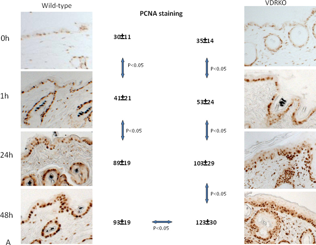
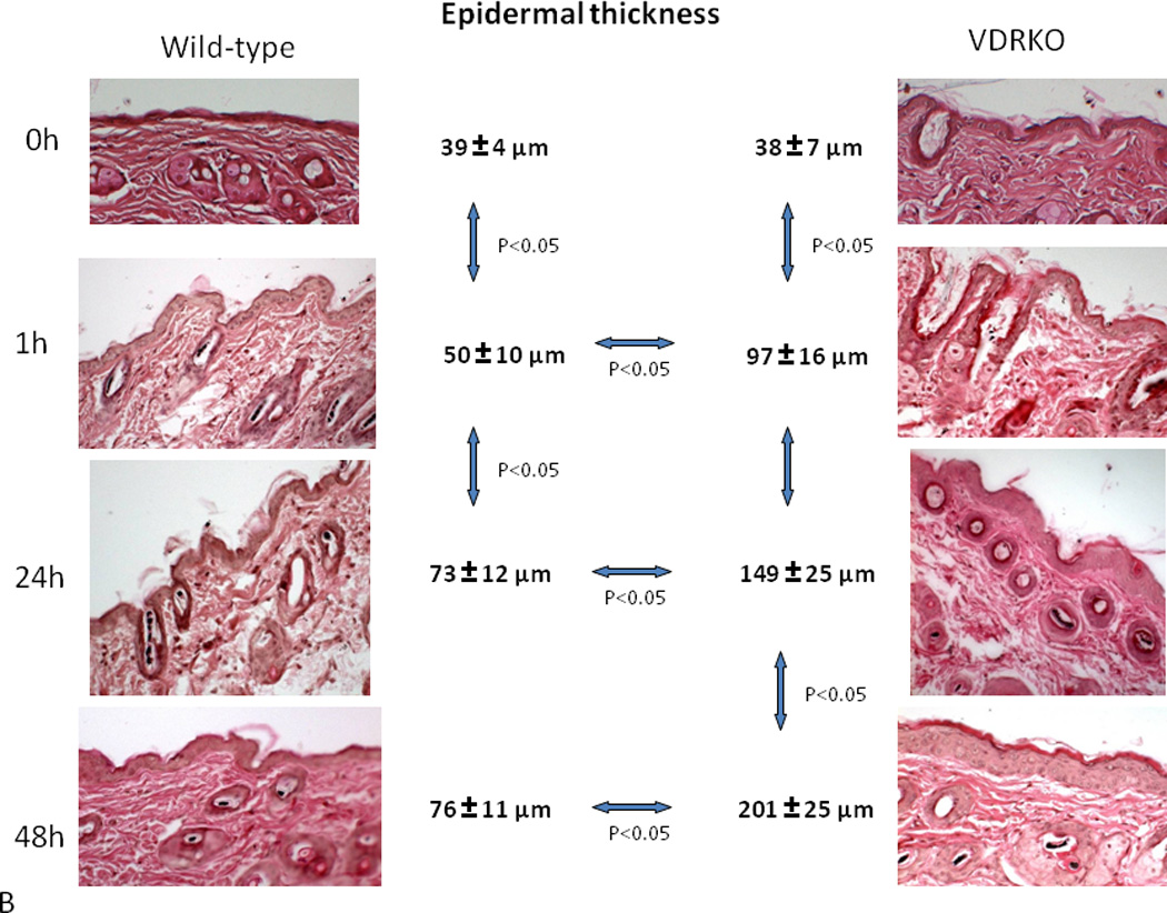
Wild-type mice exposed to 1 dose of UVB (477 mJ/cm2) showed increased proliferation (A, PCNA staining) and epidermal hyperplasia (B, H&E staining) up to 24h after treatment with no further increase at 48h. VDR null mice exposed to the same dose of UVB showed significantly more pronounced proliferation (A) and epidermal hyperplasia (B) that continued to increase at 48h. Adapted from Teichert et al. J Invest Dermatol 131:2289-2297, 2011 with permission.