
Figure 4. Defective DNA Damage Repair in VDR null mouse epidermis following UVR.
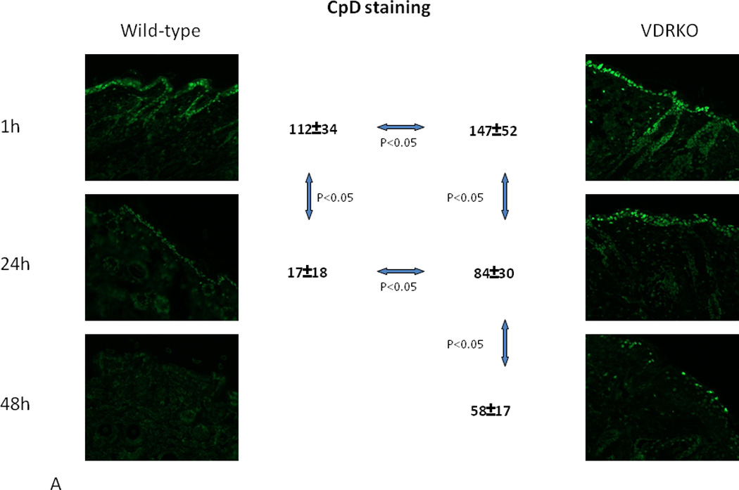
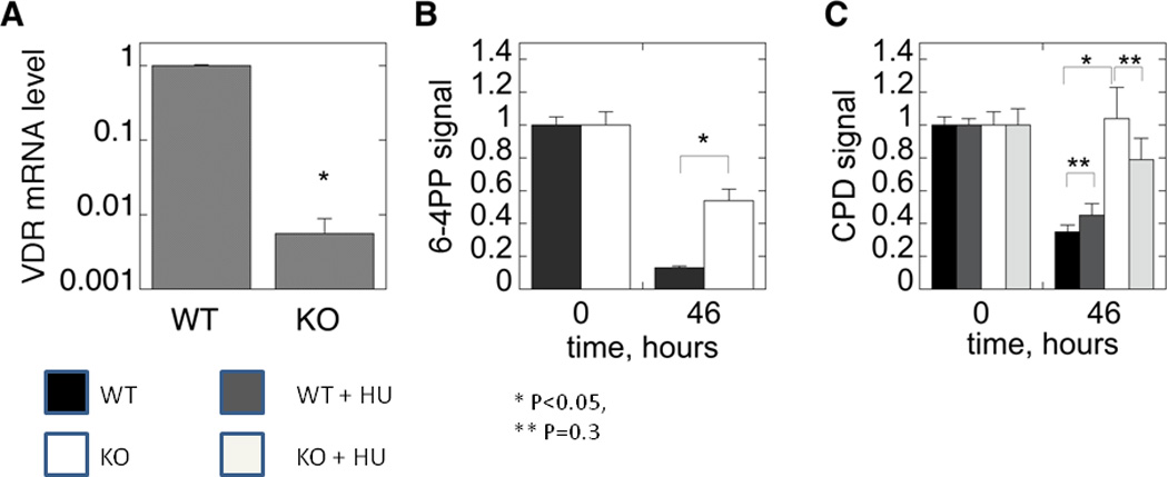
A. Wildtype and VDR null mice were exposed to 1 dose of UVB (400mJ/cm2) and the skin evaluated for the presence of CPDs over the subsequent 48hrs by immunohistochemistry (anti CPD from Cosmo Biosciences). CPDs were completely cleared by 24hr in the wildtype mouse epidermis, but persisted through 48hrs in the VDR null mouse epidermis. B. The epidermis from 2d old wildtype and VDR null mice was exposed to 35.4mJ/cm2 UVB, and CPDs and 6,4PPs (detected by immunoblots) measured immediately after irradiation and after 46hrs. In the experiment measuring CPDs, half of the epidermal explants were treated with hydroxyurea (HU) to block DNA synthesis prior to and following irradiation. Clearance of CPDs and 6,4PPs was markedly impaired in the VDR null epidermal explants consistent with the in vivo results in A. Adapted from Oh et al. J Invest Dermatol (in press, 2012).