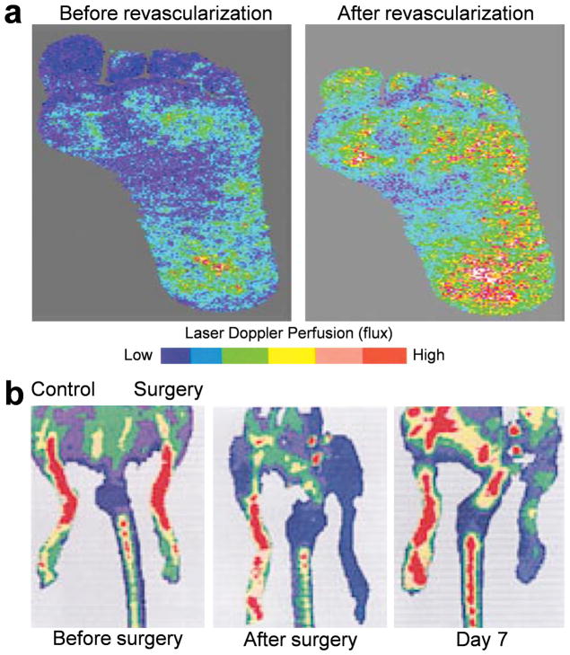
Fig. 1.
a Laser Doppler imaging of the ischemic foot of a patient with diabetes before and after infrainguinal revascularization. The areas with improved perfusion are shown in red and the areas with poor perfusion are shown in blue. b Serial laser Doppler imaging of ischemic hindlimb of a C57 mouse. Decreased perfusion soon after the surgery (dark blue) was observed in the ischemic limb, whereas high perfusion pattern (red to orange) was detected in the control hindlimb. The recovery of perfusion was clearly detectable on day 7. Adapted from [75, 76].

