
Figure 4.
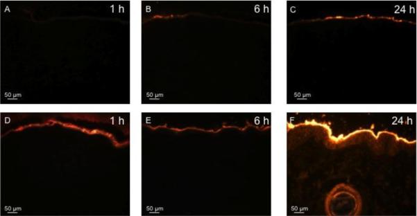
Representative histological sections of porcine skin showing permeation of red-fluorescent R-18-labeled inactivated influenza virus into skin after tissue removal by microdermabrasion. The top row shows unabraded skin at 1 h (A), 6 h (B), and 24 h (C) after applying influenza virus to the skin surface. The bottom row shows abraded skin with complete stratum corneum removal (microdermabrasion at −50 kPa, 50 passes) at 1 h (D), 6 h (E), and 24 h (F) after applying influenza virus to the skin surface. All images were collected at the same exposure time and gain using the same microscope and camera.