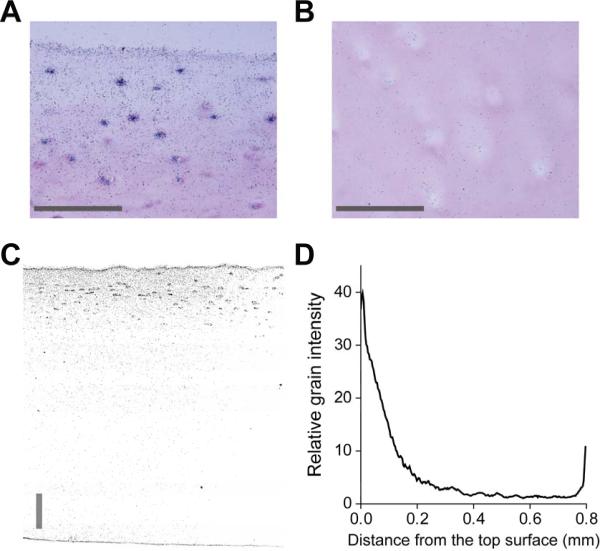
Figure 2.
Autoradiography images of 125I-Fab distributed within human knee cartilage explants at day 3. Cartilage disks were harvested from first ~ 800 μm layer of grade 0–1 human knee cartilage including the intact superficial zone. Free-swelling cartilage disks were incubated in medium containing 125I-Fab without unlabeled Fab at 37°C for 3 days in 1×PBS with 0.1% BSA, 0.01% sodium azide, and protease inhibitors. The 125I-Fab penetrated throughout the human tissue but still exhibited a pronounced gradient in grain density with depth at day 3, showing a more concentrated distribution of 125I-Fab in the upper region near the surface (A), compared to that approximately 500–600 μm below the surface (B). Distribution of 125I-Fab across the full thickness from the same disk (C) was consistent with higher magnification images (A, B), showing the gradient in the grain intensity throughout the thickness (D, relative grain intensity measured from C). Scale bars = 100 μm.

