Fig. 2.

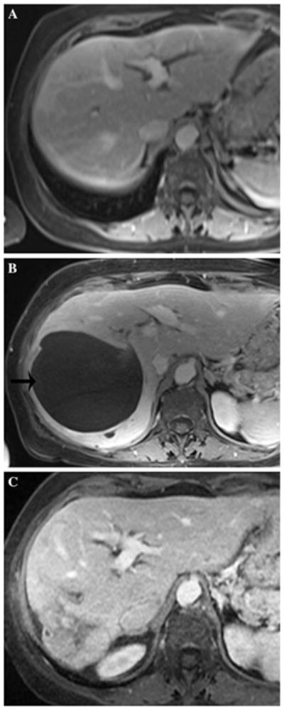
A 69 year-old woman with 70 s postcontrast T1-weighted MRI through the mid-liver showing baseline image (A), biloma adjacent to tumor after second DEB-TACE (B, black arrow on left), and resolved biloma after drainage (C)

A 69 year-old woman with 70 s postcontrast T1-weighted MRI through the mid-liver showing baseline image (A), biloma adjacent to tumor after second DEB-TACE (B, black arrow on left), and resolved biloma after drainage (C)