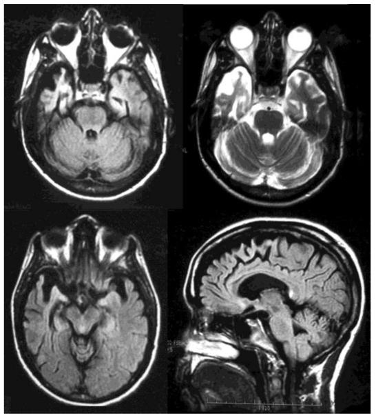
Figure 1.
Magnetic resonance imaging (MRI) of the brain for Patient No. 6. The upper left is an axial flair image showing anterior temporal areas of atrophy and gliosis, with the right side involved to a much greater degree than the left. The upper right is an axial T2 imaging of the same findings. The lower right is an axial flair image at a slightly higher level. The lower left is a saggital FLAIR image showing atrophy extending to the medial frontal region consistent with bvFTD.

