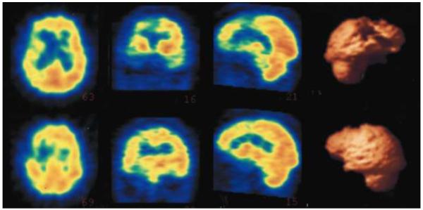
Figure 2.
Single photon emission tomography (SPECT) imaging of the brain for Patient No. 6. The images are, left to right, axial, coronal, saggital, and 3-dimensional (3-D) reconstructions of the SPECT images. They show disproprtionate hypoperfusion of the right frontotemporal region with relative sparing of the left. There is clear involvement of the right anterior temporal area extending to adjacent frontal areas. The 3-D reconstructions illustrate a lateral view of the right hemisphere in the upper right and a corresponding lateral view of the left hemisphere in the lower right.

