
Fig. 5.
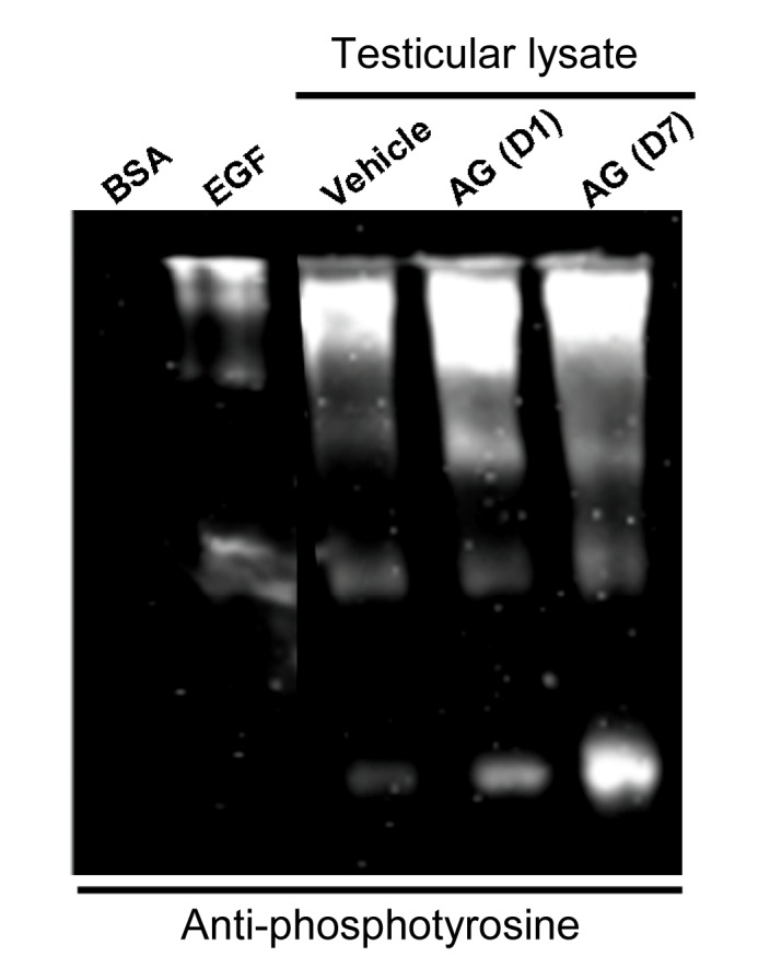
Immuno Western blot analysis for tyrosine protein phosphorylation levels in testicular lysate (50 μg for each lane) of rats treated with or without AG extract (50 mg/kg BW) on Days 1 and 7 [AG (D1) & AG (D7)]
Bovine serum albumin (BSA) and epidermal growth factor-like growth factor (EGF) were used as negative and positive controls for anti-phosphotyrosine, respectively